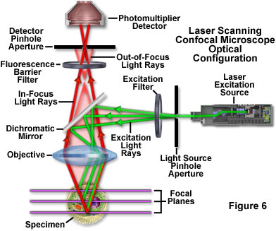
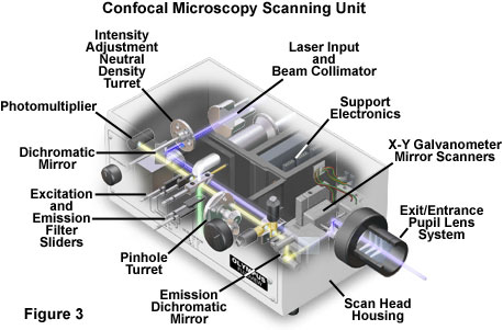
Disadvantages of confocal microscopy are limited primarily to the limited number of excitation wavelengths available with common lasers referred to as laser lines which occur over very narrow bands and are expensive to produce in the ultraviolet region.
Laser scanning microscope disadvantages.
The primary advantage of laser scanning confocal microscopy is to produce thin optical sections through fluorescent specimens that have a thickness beyond 50 micrometers.
Imaging frame rates are typically slower for single point laser.
When investigating multilayer structures the true surface of a substrate can be observed through a surface coating.
A thick section of fluorescently stained human medulla in widefield fluorescence exhibits a large amount of glare from fluorescent structures above and below the focal plane figure 1 a.
Of course you can make it faster by compromising sensitivity resolution etc and there are some special implementation of confocal such as spinning disk confocal to resolve this issue.
Electrons are reflected off the specimen to produce a 3d image.
Spinning disk systems provide an alternative means of obtaining a full frame high speed confocal image in real time.
When imaged with a laser scanning confocal microscope figure 1 d the.
In contrast conventional widefield microscopes use mercury or xenon based arc discharge lamps to provide a full range of excitation wavelengths in the ultraviolet visible and near infrared spectral regions.
A the use of microscopy to observe and investigate different types of cell and cell structure in a range of eukaryotic organisms to include an appreciation of the images produced by a range of microscopes.
The confocal laser scanning microscope s aim was not to further increase magnification but to make clearer.
The thickness of the coating can be determined by observing the 2 peaks in the axial intensity variation.
Confocal laser scanning microscopes can have a programmable sampling density and very high resolutions while nipkow and pam use a fixed sampling density defined by the camera s resolution.
A key limitation in the use of point scanning confocal microscopy for live cell imaging applications is the speed of image acquisition which may be too slow to obtain information about rapid biological processes.
The laser scans across the object and an image is built up pixel by pixel on a screen.
Laser scanning confocal fluorescence microscopy.
A typical confocal uses raster scanning which means it scans the specimen point by point.
Images are collected by coordinating incremental changes in the microscope fine focus mechanism using a stepper motor with sequential.
Laser scanning confocal microscopy.
Comparing to a wide field detection taking a snapshot of the whole field of view it is quite slow.
Advantages of confocal laser scanning microscopy industrial applications of confocal microscopy thin film profiling.


